Central Electron Microscopy Facility (CEMF)
UConn Health
UConn Health Center Research Advisory Council Cores
The Central Electron Microscope Facility (CEMF) is a UConn Health-supported research facility providing electron microscopy research service for faculty, students, and extramural users. The CEMF has three electron microscopes equipped with digital imaging capabilities available for use by experienced faculty or students.
Facility HomepageContacts
Campus Address
UConn Health
B Level: Rooms AB027, AB031
Mailing Address
UConn Health, Main Building
263 Farmington Avenue
Farmington, CT 06030
Resources
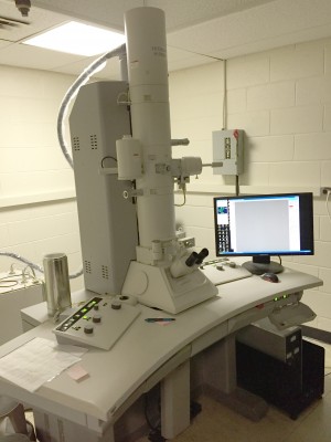
Hitachi H-7650 Transmission Electron Microscope
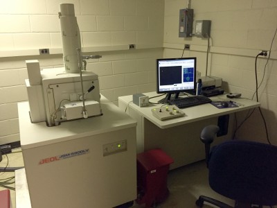
JEOL JSM-5900LV Scanning Electron Microscope
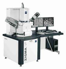
Zeiss Sigma Gemini HV Field Emission Scanning Electron Microscope
Sample Preparation Instruments
- Ultramicrotomes
- Vacuum Evaporator
- Sputter Coater
- Critical Point Dryer
- High Pressue Freezer
- Freeze Substitution System
- Ion Beam Coater
Services
TEM and SEM Specimens
CEMF offers complete processing, examination and photography of TEM (thin-sections) and SEM specimens negative staining.
Negative Staining of Particulate Samples
Other services include:
- Cryosectioning
- Freeze-fracture
- Rotary shadow
Training is available on an individual basis
List of Equipment
- Hitachi H-7650 transmission electron microscope
- JEOL JSM 5900LV scanning electron microscope (to be decommissioned in September 2023)
- Zeiss Sigma 360 variable pressure scanning electron microscope with energy dispersion spectroscopy probe (to be installed November 2023)
- Thermo-Fisher Tundra cryo-electron microscope and auto-grid assembly workstation (to be installed September 2023 in the CGSB, 400 Farmington Ave)
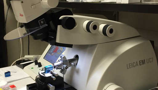
- Leica EM UC7 ultramicrotome
- Leica EM UCT ultramicrotome
- Denton Vacuum Evaporator 502B
- Denton Vacuum DESK V glow discharge unit
- Pelco Easiglow glow discharge unit
- Leica EM CPD030 critical point dryer
- 1Thermo-Fisher Vitrobot 5 (to be installed September 2023 in the CGSB, 400 Farmington Ave)