Bioscience Electron Microscopy Laboratory
Storrs
Department of Physiology and Neurobiology
The Bioscience Electron Microscopy Laboratory provides service, training and facilities required to carry out electron microscopy (EM) of a variety of biological and non-biological samples.
Facility HomepageContacts
Campus Address
Biology Physics Building, Room G06
Storrs Campus
Mailing Address
91 North Eagleville Rd., Unit 3242
Storrs, CT 06269-3242
Resources
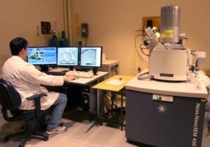
Scanning Electron Microscope (SEM)
FEI Nova NanoSEM 450: This field emission scanning electron microscope (SEM) has an ultra-stable, high current Schottky gun. Advanced electron optical and detection features include immersion mode, beam deceleration mode, and a variety of secondary and backscatter electron detectors for best selection of the information and image optimization.
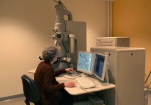
Transmission Electron Microscope (TEM)
FEI Tecnai G2 Spirit BioTWIN: This Lab6 20-120 kV transmission electron microscope (TEM) is a high-contrast, general-purpose instrument, specifically suited for low contrast samples. It enables study of low-contrast, beam-sensitive biological specimens, or other soft materials such as polymers. Samples can be either unstained or stained.
Ultramicrotomes
- Leica Ultracut UCT
- RMC MT7
- LKB Ultrotome III and V
Critical Point Dryer
Tousimis 931. GL: Critical point drying eliminates the surface tension forces that distort and collapse surfaces of hydrated samples during air drying. It is especially useful for preparing biological samples for scanning electron microscopy.
Sputter Coater
Polaron E5100: Sputter coating produces a conductive metal coating (usually gold-palladium) to reduce charging and increase secondary electron signal in the scanning electron microscope.
Microwave Tissue Processor
Pelco Biowave Pro: Accelerates fixation, dehydration, infiltration and embedding of samples for transmission electron microscopy.
Plasma Cleaner
Harrick Plasma PDC-32G: Plasma cleaning is used to remove or stabilize contaminating organic material or to make surfaces more hydrophilic for better spreading of negative stain.
CryoSEM Preparation
Leica EM VCT100, QSG100, and MED020: For freeze fracturing, freeze etching and coating SEM samples at low temperature.
Cryoultramicrotomy
Leica Ultracut UCT ultramicrotome with FCS cryo attachment: Low temperature ultramicrotomy of polymers or hydrated biological materials that cannot be sectioned at room temperature.
Freeze Substitution
Leica EM AFS: for automated freeze substitution and low temperature embedding