Mission Statement
The mission of the Center is defined by the following goals:
- To facilitate scientific discovery and theoretical and methodological innovation
- To serve as an intellectual center for interdisciplinary basic and clinical research
- To prepare graduate students and post-doctoral fellows for careers in academia and related fields
- To provide undergraduate students with research experience and other educational opportunities
- To disseminate scientific knowledge to the broader university community, relevant professional communities, and the general public
Contacts
General Contact

Nabin Koirala, Ph.D.
Director
School of Pharmacy Room 632
nabin.koirala@uconn.edu
860-486-4042

Elisa Medeiros
Technical Operations Manager
Chemistry A-316
elisa.medeiros@uconn.edu
860-486-3108
Campus Address
Phillips Communication Sciences Building (PCSB)
Storrs Campus
Mailing Address
Brain Imaging Research Center
2 Alethia Drive, Unit 1271
Storrs, CT 06269-1271
Directions & Parking
The UConn Brain Imaging Core is located in the David C. Phillips Communication Sciences Building (PCSB).
2 Alethia Drive Unit 1271
Storrs CT 06269
(860)486-3108

Two dedicated parking spaces are available for visitors in the circular lot of the David C. Phillips Communication Sciences Building. Weekdays from 7AM-5PM, please park in one of the spaces marked 'Reserved - BIRC Visitors Only'. If the reserved spots are full, park in any available spot and we will provide you with a parking pass. The David C. Phillips Communication Sciences Building is directly in front of the two parking spaces, to the right of the Human Development building.
From Route 32: Turn onto 275 East (South Eagleville Road) and follow for approximately 2 miles. At the stop light at the junction of routes 32 and 195, turn LEFT onto Route 195 (Storrs Road). Follow directions from Route 195 (below).
From Route 195: Turn onto Bolton Road (between E.O. Smith and the School of Fine Arts.) Take your second RIGHT onto Alethia Drive, and then your second LEFT into the circular parking lot. Park in one of the reserved spaces.
You can print a copy of these directions.pdf

Campus Map and Information Enter "PCSB" in the "search the map" box
Researchers: Please use the template directions for research participants.docx rather than directing them to this page.
Instrumentation
Laboratory
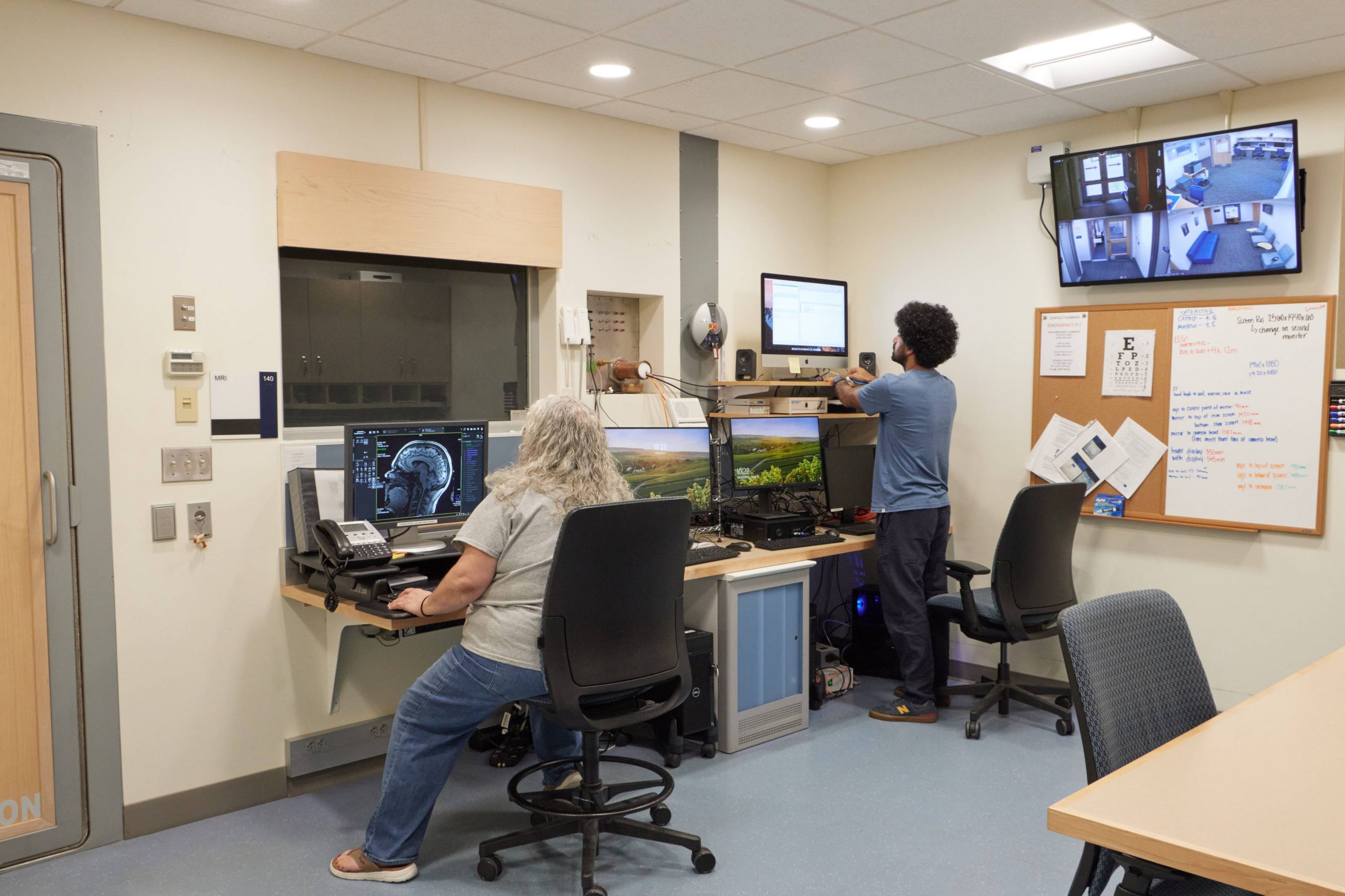
The BIRC facilities include dedicated spaces for functional brain imaging for computing, and image and data analysis. The center thus contains all of the resources necessary for integrated studies of human brain function. It houses all of the research scanners and personnel in one contiguous facility.

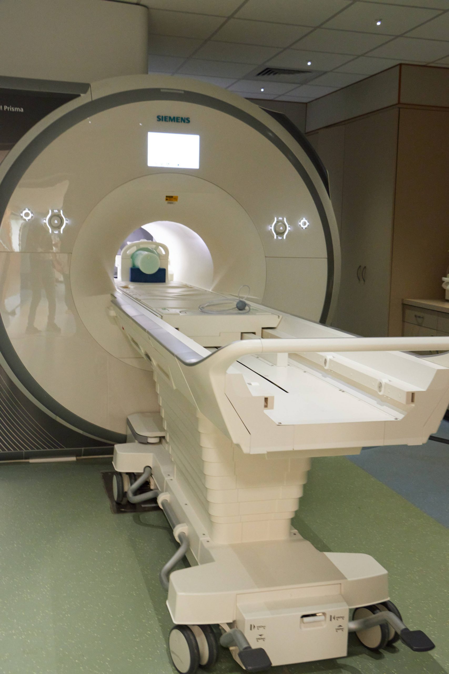
MRI 3.0 Tesla Siemens Prisma Scanner
A 3.0 Tesla Siemens Prisma scanner is available for structural and functional MRI scanning, as well as for magnetic resonance spectroscopy (MRS). This system has 80mT/m maximum amplitude gradient hardware, and a slew rate of 200T/m/sec with a 100% duty cycle. It also includes 20- and 32-channel head and 64-channel head/neck receive array coils with parallel imaging capabilities. This system is also equipped with 32-channel spine coil, shoulder coil, hand/wrist coil, foot/ankle coil, knee coil, and 18-channel body coil. The signal quality and operating characteristics of the scanner are routinely monitored using ACR, fBIRN, and dynamic phantoms.
Mock MRI: There is also an MRI simulator in a dedicated room in the BIRC that allows for training of subjects that might have difficulty staying still in the magnet (such as children) and also for subjects that might suffer from mild claustrophobia. The mock scanner includes equipment for displaying stimuli and for monitoring and providing feedback on head motion via and will be used for orientation to the MRI environment and training and in-scanner tasks.

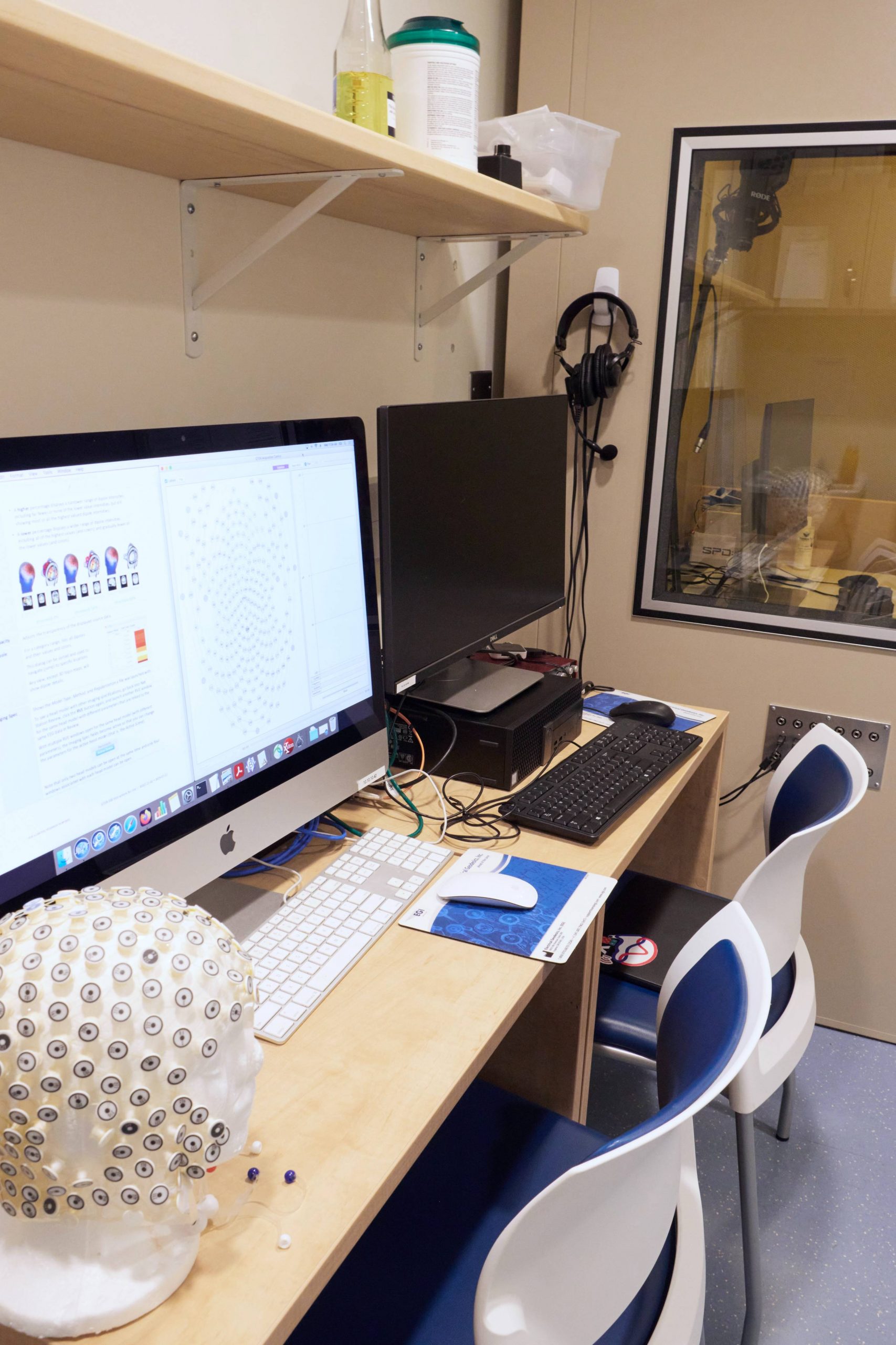
EEG: 256-Channel EEG System (EGI)
The BIRC houses two 256-channel EEG systems from EGI with a full range electrode caps in pediatric and adult sizes. A Net Amps 410 is available for simultaneous EEG/MRI and a Net Amps 400 GTEN system is available for out-of-scanner use. The latter is equiped with a neuromodulation package and software for high density tDCS, tACS, tRNS and tPCS. An EGI Geodesic Photogammetry System (GPS) is available for electrode localization in conjunction with EEG or tDCS studies.

Computing
The center houses a high-end data processing lab, featuring 4 high-end Mac Workstations with common software (e.g. Docker, AFNI, FSL, Freesurfer, MATLAB, R). In addition to in-house computing, two high performance computing (HPC) systems are available to UConn faculty, providing access to accelerated GPU and parallel computing. BIRC has purchased one semi-dedicated 32-core processing node for priority access.
High-Performance Computing (HPC) facilities
The University manages HPC systems on the Storrs campus and in Farmington at UConn Health. The systems are optimized for different computational problems, with the Storrs focusing on compute-intensive workloads and the Farmington facility on data-intensive workloads. Both provide access to accelerated GPUs, parallel computing, and 1 PB of parallel file storage and 3 PB archival storage distributed across multiple datacenters. The Farmington facility has 3,000 processor cores, and the Storrs HPC cluster currently has over 11,000 CPU cores on 400 nodes. We maintain a data transfer node with Globus software for large data transfers both within and outside of the environment. Network traffic travels over Ethernet at 10Gb per second between nodes, and file data travels over InfiniBand at 56Gb or 100Gb per second, depending on the node. All UConn researchers may use the community queues, but researchers may purchase semi-dedicated nodes for high priority access. Computational applications, including containers, are installed as needed; to date, over 200 have been made available.
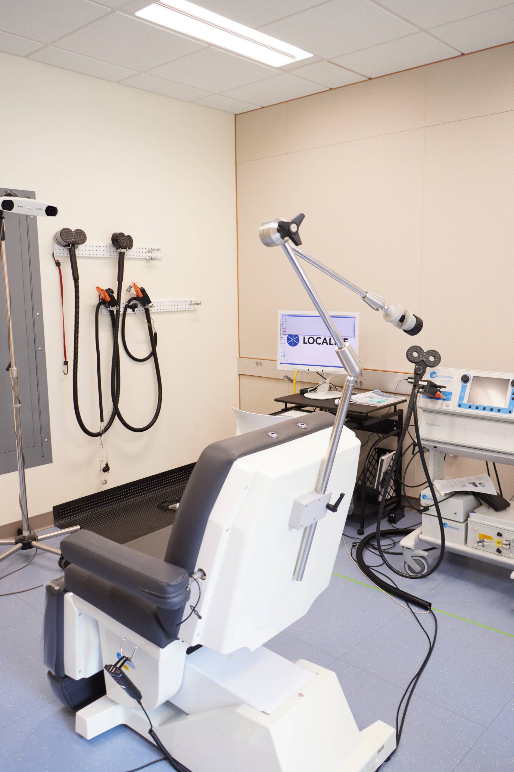
MagVentures Transcranial Magnetic Stimulation (TMS)
A MagVentures MagPro X100 with MagOption is available for TMS. This system is capable of generating high duty biphasic and monophasic pulse sequences for single pulse, paired pulse or repetitive stimulation. The system is equipped with flat and 120º butterfly coils, and a liquid-cooled blinded active/sham butterfly coil. Pulse localization on individual anatomy is facilitated by a Localite TMS Navigator system.
