Announcements & Events
The new Nikon AXR Confocal + TIRF instrument is up and running. It offers fast and sensitive confocal imaging and also has a camera for other modalities such as TIRF, transmitted light, and epifluorescence imaging. There also also a range of denoising and deconvolution options for processing images. Use can be arranged by filling out the Service Request Form.
Microscope Reservation Policies
- Microscope reservations can be made no more than 14 days in advance.
- Reservations can be made in 15 min increments starting on the quarter hour.
- There is no maximum reservation time limit.
- Changes or cancellations to reservations can be made up to 6 hours in advance. After that, the full cost of the reservation will be charged.
- The actual reservation time plus any additional time used will be billed automatically.
- Reservations will be automatically cancelled if not started within 30 min.
- Once per user, per calendar month, a billing adjustment can be made for a cancelled/changed reservation. Requests must be made to Chris O’Connell by email
Service Requests
Assisted sessions and instrument training can be requested by filling out the Service Request Form. Rates for instruments and service can be found here. After your form is received, you will be contacted to arrange a brief meeting to discuss your projects imaging needs to determine whether assisted sessions or training are necessary, the best instrument for your application, and any sample preparation considerations. Refer to the below flowchart which outlines the process for using the facility.
Staff
Christopher O'Connell, Ph.D.
Facility Director
Biology Physics Building, Rm G05C
coconnell@uconn.edu
860-486-3271
CJenna Martin, Ph.D.
Facility Scientist
Biology Physics Building, Rm G05A
jmartin@uconn.edu
860-486-4550
Faculty Scientific Advisers

Juliet Lee, Ph.D.
Associate Professor
Biology/Physics Building 306
juliet.lee@uconn.edu
860-486-4332

Akiko Nishiyama, Ph.D.
Professor
Pharmacy/Biology Building 631
akiko.nishiyama@uconn.edu
860-486-4561
George Lykotrafitis, Ph.D.
Associate Professor
UT Engineering Building Rm. 374
george.lykotrafitis@uconn.edu
860-486-2439
Campus Address
Biology Physics Building, Room G05D
Mailing Address
Advanced Light Microscopy Facility
Center for Open Research Resources and Equipment
75 North Eagleville Road, Unit 3149
Storrs, CT 06269
Instrumentation
Leica SP8 Spectral Confocal
This is an inverted confocal microscope with five filter-free spectral and individually regulatable channels including four standard PMT detectors and one high-sensitivity HyD detector with photon counting capacity. The scanner also includes both standard galvo scan mirrors and an optional 8 KHz resonant scanner for high speed imaging. Use of an acusto-optical beamsplitter (AOBS) instead of traditional dichroic mirrors makes this microscope also suitable for reflected light microscopy applications. The instrument is housed in the Bioscience Electron Microscopy Laboratory located in the Biology and Physics Building (BPB), room G10.
Specifications
Location:
Bioscience Electron Microscopy Laboratory
Biology and Physics Building (BPB), room G10.
Specifications:
Microscope:
- Inverted Stand DMI 6000
- Motorized stage
- Super Z Galvo stage
Lasers:
- 405 nm Diode laser
- Argon/2 (458, 488, 514 nm)
- 561 nm DPSS laser
- 633 nm HeNe laser
Scanners:
- Continuously adjustable non-resonant scanner (1-1800 Hz)
- Resonant scanner (8000 Hz) – 28 fps at 512 x 512
Objective Lenses:
- 10x/0.30 HC PL FLUOTAR
- 20x/0.75 HC PL APO IMM CORR CS2
- 40x/1.30 HC PL APO OIL CS2
- 63x/1.40 HC PL APO OIL CS2
- 100x/1.40 HCX PL APO OIL CS
Detectors:
- Four PMTs
- One HyD
- All filter-free spectral and individually regulatable detectors
- Transmitted light brightfield detector (with DIC option)
Software:
- LAS X for image acquisition, processing, and quantification
Internal Rates
$21.60/hour Unassisted Use
$88.60/hour Assisted Use
$88.60/hour Instrument Training
External Rates
Please contact for rates and information.
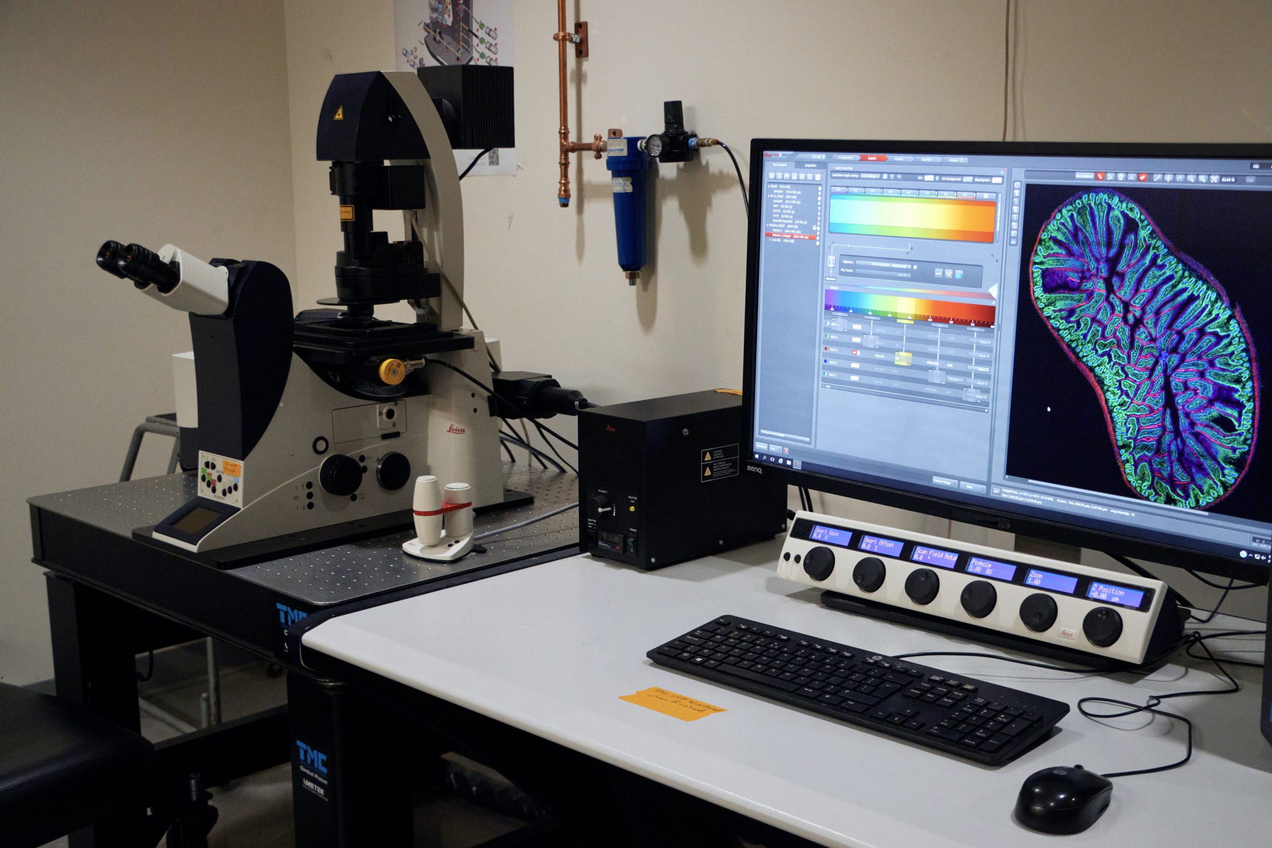
Nikon AXR Confocal + TIRF
The Nikon AXR is a point scanning confocal built around the inverted Ti2E microscope. The large 25 mm field of view and resonant scanner are optimized for rapid imaging of samples. The DUX-VB confocal detector features 4 sensitive GaAsP detectors that can be flexibly configured for a range of dyes via spectral detection. 6 laser lines provides a range of excitation options for a variety of fluorescent dyes. In addition to confocal microscopy, the system has an iLAS2 illumination module for TIRF imaging. This device can perform simultaneous TIRF imaging and photostimulation. TIRF, widefield, and transmitted light images are collected on a Photometrics Prime 95B sCMOS camera. The instrument is located in the Biology and Physics Building (BPB), room G05D.
Specifications
Specifications:
Microscope:
- Inverted microscope Nikon Ti2E
- Motorized x/y stage
- PerfectFocus optical z drift correction
- MCL Nano-Z200 piezo z stage
Laser Unit for Confocal:
- LUA-S6 405/445/488/514/561/640
Laser Unit for TIRF:
- LUNF XL 405/488/561/640
TIRF/Widefield Camera:
- Photometrics Prime95B
Objective Lenses:
- 10X PLAN APOCHROMAT LAMBDA D, DIC
- 20X PLAN APOCHROMAT LAMBDA D, DIC
- 40X/1.30 PLAN FLUOR, Oil, DIC
- 60X/1.40 PLAN APOCHROMAT LAMBDA D, Oil, DIC
- 100X/1.45 PLAN APOCHROMAT, Oil, DIC
- 100X/1.49 APO TIRF, Oil, DIC
Software:
- NIS-Elements C Confocal Package, JOBS for Scanning Plates and Creating Custom Acquisition and Analysis Routines, NIS-Elements 2D/3D, Deconvolution Suite, NIS.ai Artificial Intelligence Module, General Analysis 2 and 3
Internal Rates
$21.60/hour Unassisted Use
$88.60/hour Assisted Use
$88.60/hour Instrument Training
External Rates
Please contact for rates and information.
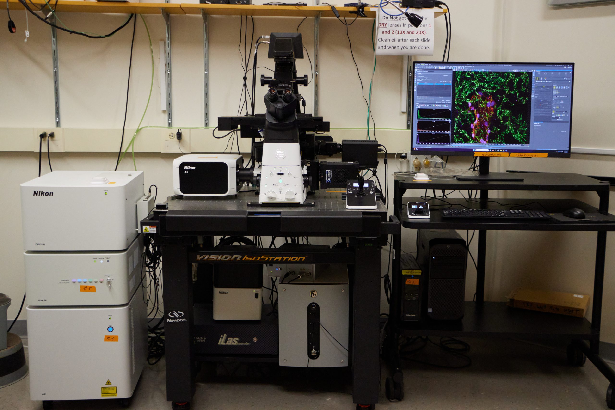
Image Analysis Workstation
The Image Analysis Workstation is a powerful computer dedicated for image analysis and visualization. Several software packages are installed to support a variety of data formats.
Specifications
Location:
Bioscience Electron Microscopy Laboratory
Biology Physics Building (BPB), room G10.
Specifications:
Hardware:
- HP Z640 Workstation
- 30″ Z30i LED-LCD Monitor
- Microsoft Windows 7 Professional Edition 64bit OS
- NVIDIA Quadro K2200 4GB Graphics Card
- Intel® Xeon E5-1660v3 3.00GHz 20MB 2133 8C CPU
- 64GB RAM
- 2 TB striped RAID data drive
Software:
- Leica LAS X including the following modules:
- 2D Analysis, 3D Analysis, and 3D Visualization
- NIS-Elements AR including the following module:
- Deconvolution
- Image J
Internal Rates
No charge for Confocal Microscopy Facility Users.
A Google calendar is maintained for reserving time on the workstation.
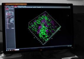
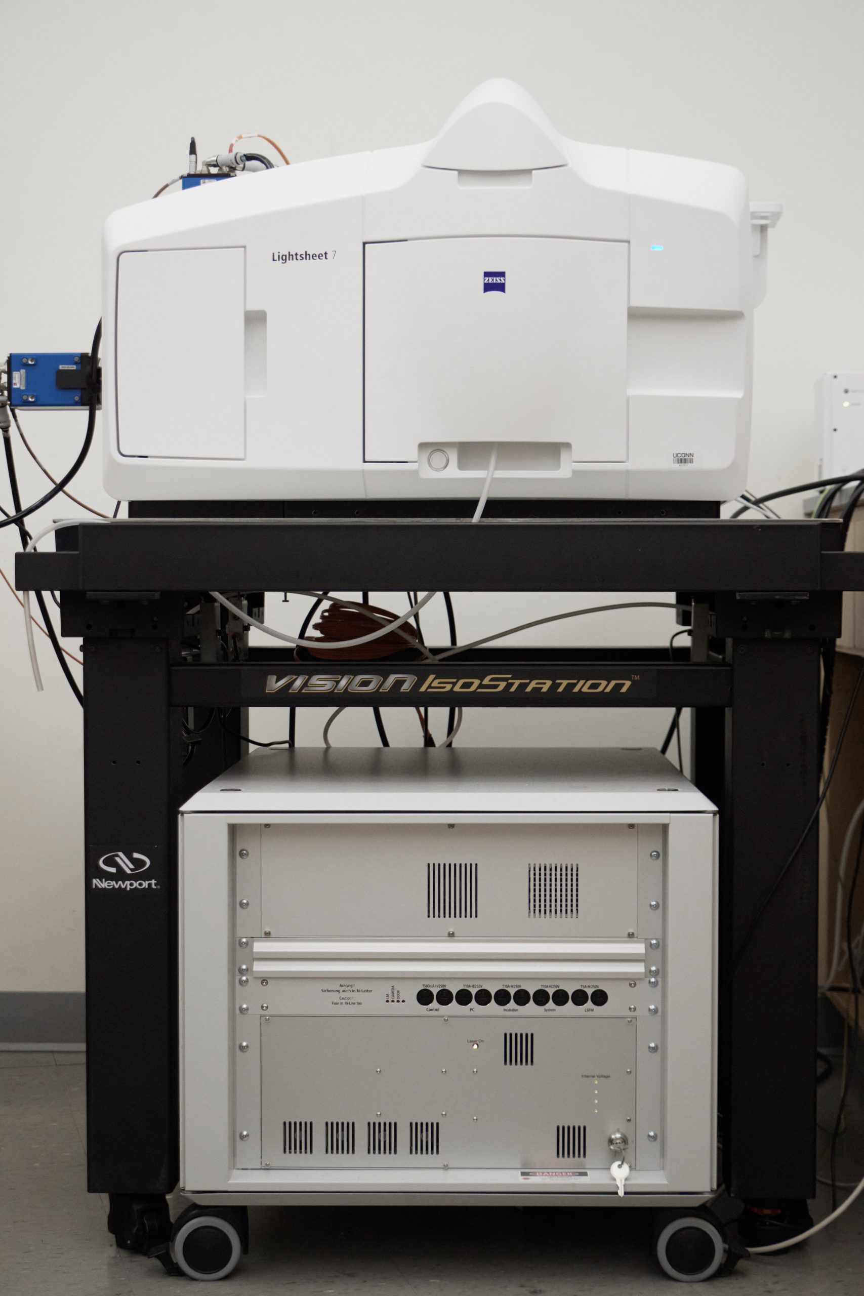
Zeiss Lightsheet 7
The Zeiss Lightsheet 7 microscope is designed for fast, gentle 3D/4D imaging of samples labeled with fluorescent probes. Both live and fixed samples can be imaged. Samples are lowered from above into an imaging chamber containing media of the appropriate refractive index. It is located in BPB G05.
Specifications
Lasers:
- 405, 488, 561, 638 nm
Imaging Optics:
- 5X 0.16NA (for live samples in aqueous media)
- 5X 0.16 NA (for fixed samples, correction collar adjustable for different refractive index media up to RI=1.53)
- 10X NA 0.5 (for live samples in aqueous media)
- 20X NA 1.0 (for live samples in aqueous media)
- 20X NA 1.0 (for fixed samples, correction collar adjustable for different refractive index media up to RI=1.45)
Illumination Optics (2 each for forming the lightsheet):
- 5X NA 0.1
- 10X NA 0.2
Imaging Chambers:
- Live chamber for samples in aqueous media
- Fixed sample chamber (small)
- Fixed sample chamber (large, 28 x 31 x 49 mm)
Detectors:
- 2 PCO edge 4.2 sCMOS cameras for dual channel imaging
- 2048 x 2048 pixel resolution
- 100 fps maximum frame rate
- up to 82 % quantum efficiency
Software:
- Zen Black for acquisition
- Zen Blue for analysis
- Arivis for large volume 3D rendering and analysis (on separate analysis PC)
Internal Rates
$17.40/hour Unassisted Use
$84.40/hour Assisted Use
$84.40/hour Instrument training
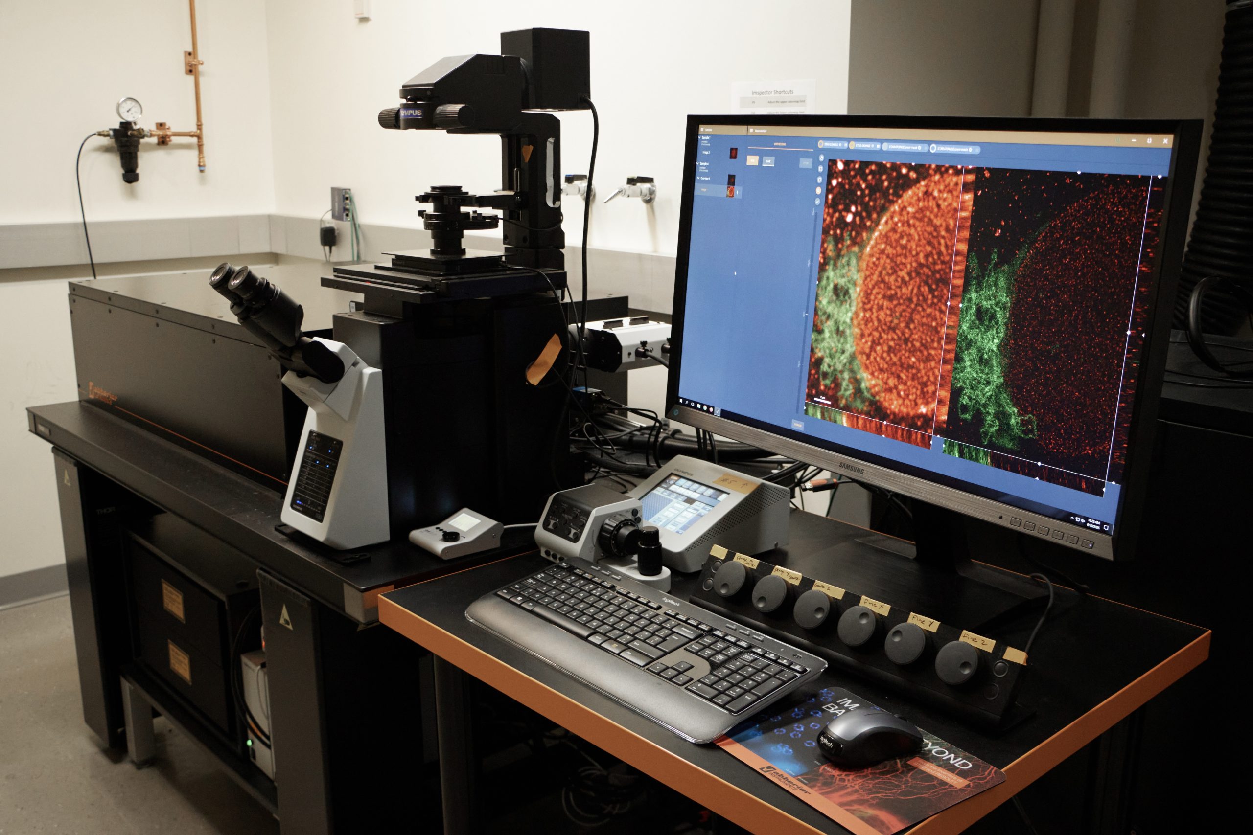
Abberior Instruments 3D STED
This is an inverted confocal microscope that uses stimulated emission depletion (STED) of fluorescent dyes to obtain super resolved images. There are four pulsed excitation lasers and two pulsed STED depletion lasers for imaging. The instrument is capable of 2D or 3D super resolution imaging using spatial light modulators to shape the depletion beams in the x-y and z axes. The instrument is housed within the Bioscience Electron Microscopy Laboratory located in the Biology and Physics Building (BPB), room G11.
Specifications
Microscope:
- Inverted Stand Olympus IX83
- Motorized x-y stage
- Physik Instrumente piezo z stage insert
Lasers:
Excitation
- 405 nm CW (non-STED imaging)
- 440 nm pulsed
- 485 nm pulsed
- 561 nm pulsed
- 640 nm pulsed
STED Depletion
- 595 nm pulsed
- 775 nm pulsed
Scanner:
- Continuously adjustable QUADScan galvo scanner (up to 2600 Hz line scanning)
Objective Lenses:
- 10X/0.3 UPLFLN10X2
- 20x/0.75 UPLSAPO20
- 40x/0.1.3 UPLFNL40 oil immersion
- 60x/1.2 UPLSAPO60 water immersion
- 100x/1.40 UPLANSAPO oil immersion
Detectors:
- Four filter-based APD detectors
Software:
- Abberior Impsector for acquisition and visualization
Internal Rates
$22.80/hour Unassisted Use
$89.80/hour Assisted
$89.80/hour Instrument training
Leica Thunder Imager
The Leica Thunder Imager is an upright microscope for fluorescence, transmitted light, and true color imaging . With a stage capable of holding up to 8 slides, this instrument is optimized for high throughput imaging of cells and tissue sections. Fluorescent images can be processed with Leica’s computational clearing and deconvolution to remove out of focus blur, enhance contrast, and sharpen details. The software Navigator function can be used for rapidly generating sample overviews for subsequent selection of areas of interest. The instrument is located in the Biology and Physics Building (BPB), room G05D.
Specifications
Internal Rates
$11.40/hour Unassisted Use
$78.40/hour Training and Assisted Use
Services and Rates
The Advanced Light Microscopy Facility provides training and access to advanced light microscopy systems at an hourly rate. In addition, we are available to consult with and support users at every stage of a project including: experimental design, sample preparation, image acquisition, analysis, and data preparation.
- Consultation: We are available to meet with current and potential users to advise on sample preparation and the selection of the instrument that best serves the research requirements.
- Training: All users are provided with training that covers facility policies and details of instrument operation.
- Instrument Access: Researchers are able to independently use the instruments after training, or they can work with the Director in assisted sessions. See Instrumentation for current rates.
- Support: Our expertise is always available to help maximize the impact of the instruments on users’ studies through further consultations or by scheduling assisted sessions with the Director on the microscope.
- Education: The Facility gives workshops and lectures on microscopy applications and image analysis to advance the microscopy knowledge of the research community.
- Billing: Monthly usage is tabulated and billing is executed by the Center for Open Research Resources and Equipment (COR2E).
Microscopy Resources
Fluorescence Spectra Viewers
ThermoFisher SpectraViewer
FPbase
Microscopy Software
Fiji (ImageJ bundled with useful plugins)
Bioformats Plugin
ImageJ Forum