Services
Magnetic Resonance Imaging (MRI)
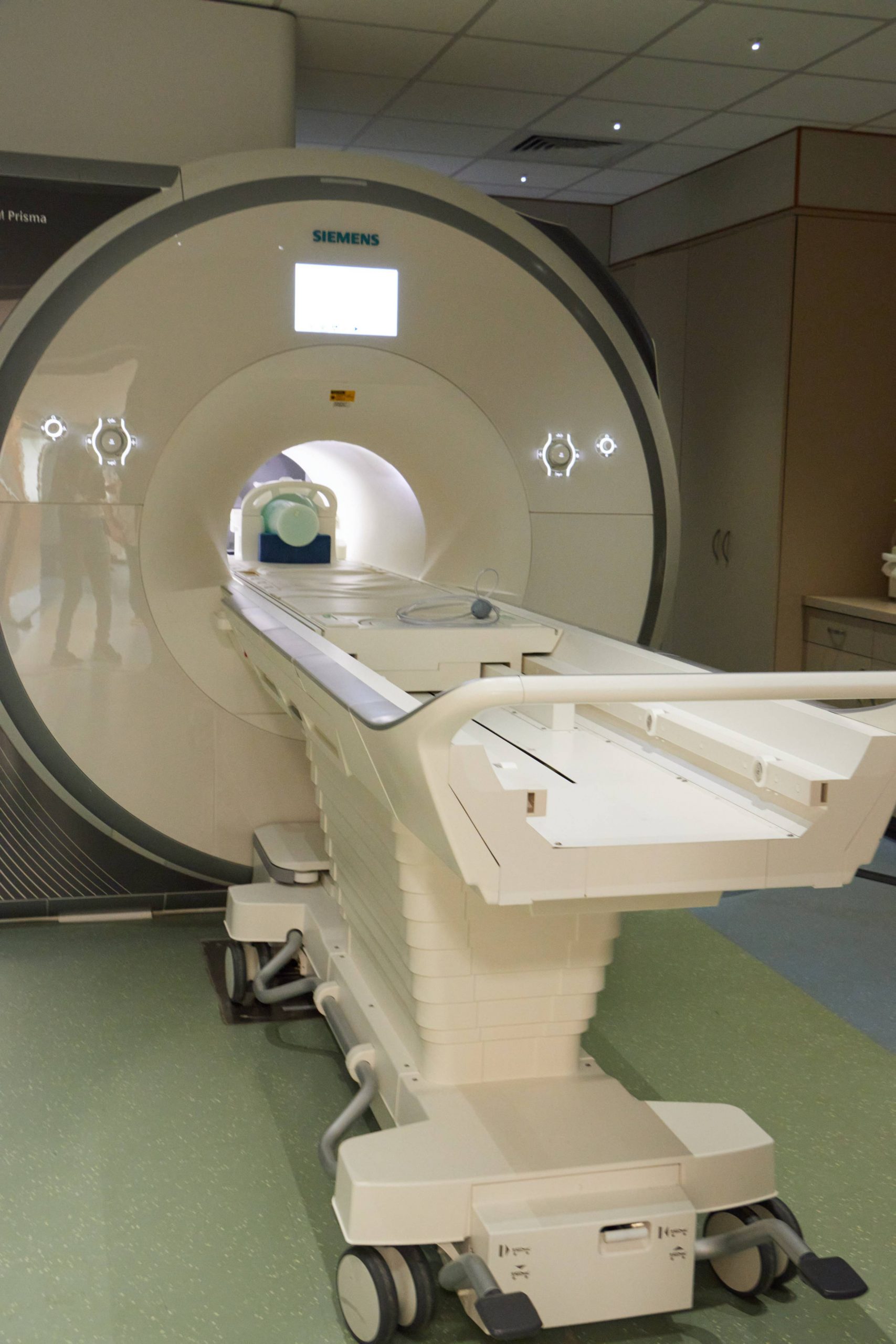
A 3.0 Tesla Siemens Prisma scanner is available for structural and functional MRI scanning, as well as for magnetic resonance spectroscopy (MRS). This system has 80mT/m maximum amplitude gradient hardware, and a slew rate of 200T/m/sec with a 100% duty cycle. It also includes 20- and 32-channel head and 64-channel head/neck receive array coils with parallel imaging capabilities. This system is also equipped with 32-channel spine coil, shoulder coil, hand/wrist coil, foot/ankle coil, knee coil, and 18-channel body coil. The signal quality and operating characteristics of the scanner are routinely monitored using ACR, fBIRN, and dynamic phantoms.
Specifications
BIRC houses a 3-Tesla Siemens Prisma scanner equipped with an eye-tracker as well as a range of head and body coils:
- 20-channel head/neck coil
- 32-channel head coil
- 64-channel head/neck coil
- 32-channel spine coil
- Large and Small Shoulder coils
- Large and Small Flex coils
- Hand/Wrist coil
- Foot/Ankle coil
- Knee coil
- 18-channel body array coil
Product and research pulse sequences for:
- Structural imaging (Siemens or MGH sequences)
- Diffusion imaging (Siemens or CMRR sequences)
- BOLD
- Susceptibility Weighted Imaging (SWI)
- Spectroscopy and chemical shift imaging (Siemens or CMRR sequences)
- Simultaneous Multislice (SMS) EPI (Siemens or CMRR sequences)
- Arterial Spin Labeling
Stimulus presentation and response hardware for high fidelity auditory and visual presentation:
- Avotec Silent Scan audio system
- Optoacoustics OptoACTIVE II audio system
- Hyperion MRI Digital Projection System
- Current Designs MR-compatible response pads
- In-Scanner Eye Tracking (SR Research Eye Link 1000 Plus)
- Presentation using a PC, Mac, or your own laptop
Mock MRI
Mock MRI: There is also an MRI simulator in a dedicated room in the BIRC that allows for training of subjects that might have difficulty staying still in the magnet (such as children) and also for subjects that might suffer from mild claustrophobia. The mock scanner includes equipment for displaying stimuli and for monitoring and providing feedback on head motion via and will be used for orientation to the MRI environment and training and in-scanner tasks.
MRI Training
Investigators and students wishing to conduct research using our Siemens Prisma 3T scanner will need to complete Advanced Safety Training at BIRC. This involves a period of approximately 90 minutes, during which you will receive hands-on orientation to the MRI suite (Zones 3 and 4). Note: you must complete Level 1 safety training and complete all required reading (provided when AST session is scheduled) prior to the session. Please contact Elisa Medeiros to schedule safety training.
MRI Safety and Screening
Participant Safety
All participants need to complete a Metal Screening Form.pdf prior to participating in a MRI. We recommend having the form completed prior to the participant’s appointment day so any potential contraindications can be addressed. The form will be reviewed by the scanner operator at the time of the MRI. The researcher should retain the signed form, consistent with their IRB-approved data retention policy. Below is a list of common issues presented by participants:
- Permanent retainers: Most are MRI compatible, and create only a small artifact that does not affect data collection; however, some retainers are mildly attracted to the magnet. Unfortunately there is no way to know if the retainer is attracted or not other than to have the participant come in and be tested by entering the magnet room with the technologist. Please note that some retainers that were safe during previous MRIs done on a 1.5T scanner may be attracted to a 3T scanner.
- Braces: While braces are not a contraindication for MRI, the artifact that is created obliterates anatomy in the fMRI and diffusion sequences; therefore, consider excluding any participant that has braces.
- Colored contact lenses: Some pigments used in colored contacts contain iron oxide and are not MRI safe. Clear contact lenses can be worn, and MRI compatible glasses can be provided.
- Tattoos: A small percentage of tattoos can be affected during a MRI resulting in a burn. We suggest avoiding having a study done until the tattoo is fully healed (approximately six weeks).
- Clothing: All participants are required to change into BIRC scrubs. Lockers are provided for personal belongings.
We also have information for participants that can help them prepare for a study.
Scanner QA
The scanner has been extensively validated in internal tests. We have replicated multiple visual field studies as well as functional localizers across scanners from multiple other sites. We perform QA tests every week using both ACR (American College of Radiology) and Siemens phantoms. In addition, we perform a scanner stability measurement from MGH. Results from these three weekly tests are automatically processed by routines at the BIRC and are monitored carefully for any sudden changes in the output. The scanner is remarkably stable, which we have attributed to both our diligent efforts at managing the magnetic field as well as regular service from Siemens.
In-scanner eye-tracking
Eyelink 1000 Plus
The UConn Brain Imaging Research Center (BIRC) houses an Eyelink 1000 Plus eye tracker, which is the fastest, most accurate, and most precise eye tracker for MRI environments.
Eye-tracking during scans allows researchers to monitor participants’ eye movements while simultaneously collecting fMRI data, and subsequently employ advanced analytical techniques such as fixation-related (FIRE) fMRI.
The eye tracker can be used with multiple stimulus presentation software including Experiment Builder, PsychoPy, E-Prime, NBS Presentation, Psychtoolbox, and OpenSesame.
If you are interested in adding eye tracking as an additional measure to fMRI, please contact gitte.joergensen@uconn.edu.
Dr. Joergensen has many years of eye-tracking experience and will provide support with how to best integrate eye tracking with fMRI, as well as other aspects of eye-tracking.
EyeLink 1000 Plus Long Range Mount MRI Setup and Usage Training Videos (SR Research)
System Overview – This video provides an overview of the EyeLink 1000 Plus Long Range Mount for MRI
Mirror Replacement -This video provides the basics of first surfaced mirror for eye tracking in MRI and how to handle and identify a first surface mirror.
Screen Mount (Horizontal Positioning) – This video provides an overview of setting up a Screen Mount (Horizontal Positioning)
Screen Mount (Vertical Positioning) – This video provides an overview of setting up a Screen Mount (Vertical Positioning)
Eye Tracker Positioning – This video provides an overview to finalize the position of the EyeLink 1000 Plus Long Range Mount.
Participant Setup – This video provides an overview to setup and calibrate a participant.
Drift Check / Correct Options – This video provides an overview of Drift Check and Drift Correct options.
Visual Angle Calculator (SR Research) – Visual angle typically refers to the angle a visual stimulus subtends on the eye.
Other resources: Recorded webinars of MRI-related courses and talks
- Coursera – Princicples of MRI 1 (see other courses as well)
- MIT fMRI Bootcamp
- Computational Psychiatry Course at TNU in Zurich
- Organization for Human Brain Mapping (OHBM)
- UCLA summer imaging course (2015)
- Brain Space Initiative
- Dartmouth Cognitive Neuroscience Speaker Series
- ENIGMA-U – Neuroscience School for high school/undergraduates
*If you have other good resources to add here, please contact us.
Electroencephalography
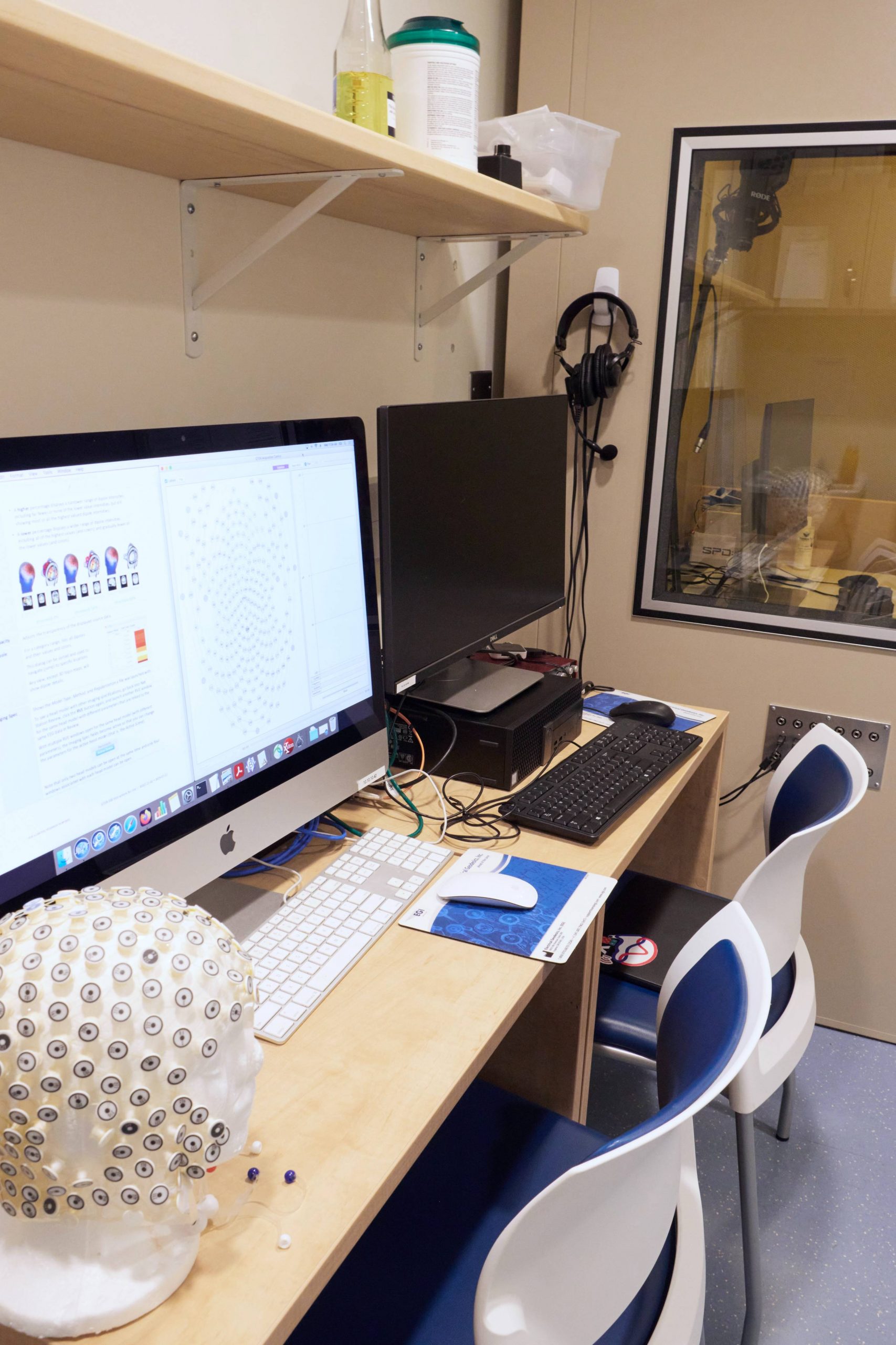
EEG: 256-Channel EEG System (EGI)
BIRC houses two 256-channel EEG systems from EGI with a full range electrode caps in pediatric and adult sizes. A Net Amps 410 is available for simultaneous EEG/MRI and a Net Amps 400 GTEN system is available for out-of-scanner use. The latter is equiped with a neuromodulation package and software for high density tDCS, tACS, tRNS and tPCS. An EGI Geodesic Photogammetry System (GPS) is available for electrode localization in conjunction with EEG or tDCS studies.
Specifications
BIRC houses two high-density (256-channel) Philips/EGI gel-free EEG systems. A 64-channel Brain Products actiCap system is available at CSSERL.
- 256-channel EGI NetAmps 400 EEG system for out-of-scanner use
- MR-compatible 256-channel EGI NetAmps 410 for simultaneous MR-EEG recording
- Sound-attenuated booth for EEG and behavioral experiments with visual and auditory presentation
- EGI Geodesic Photogrammetry System for electrode localization
- Complete range of electrode net sizes for child and adult friendly electrode application
- Physio16 box (MR compatible) for recording ECG or other physiological signals
- Cedrus StimTracker for precise event marker timing
The EGI NetAmps 400 is also equipped with the GTEN neuromodulation package for high-definition transcranial electrical current stimulation (tDCS/tACS/tPCS).
EEG Training
EEG training is required prior to using the EGI high density EEG systems at BIRC. In-scanner users must also complete MRI training. Training consists of an orientation session and at least three supervised practice sessions. Please complete the BIRC EEG Training Request Form. Researchers should also complete EHS Biological Safety Considerations: Human Subjects Research (HuskyCT) training. Researchers are strongly encouraged to operate the EEG system in pairs. You will receive card access to the EEG room after completing training.
EEG Safety and Screening
Participants with open head wounds must be excluded from EEG studies. Since the EGI system does not use gel, some hairstyles (bald or partially bald, very short, tightly braided, or very springy hair) will make obtaining good electrode contact with the scalp difficult or impossible. Certain hair dyes, generally reddish hues, may bleed in contact with the saline solution. You should inform the participant of this and get permission to test the dye by applying a small amount of saline solution to a patch of non-visible hair.
MagVentures Transcranial Magnetic Stimulation (TMS)
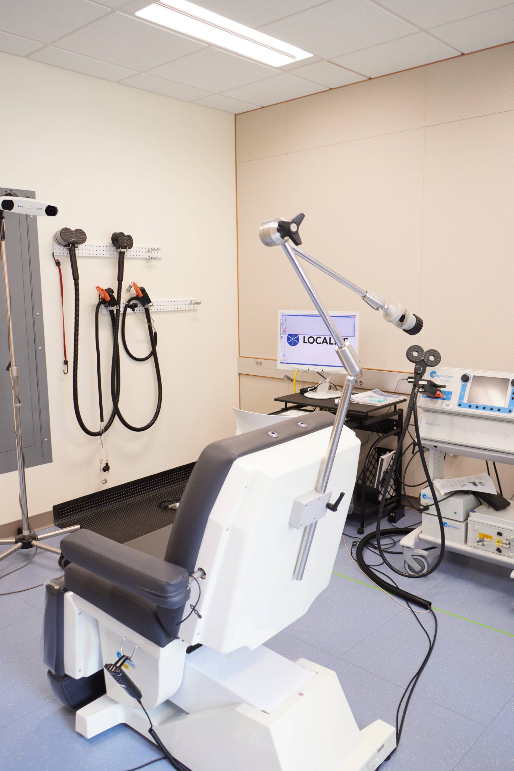
A MagVentures MagPro X100 with MagOption is available for TMS. This system is capable of generating high duty biphasic and monophasic pulse sequences for single pulse, paired pulse or repetitive stimulation. The system is equipped with flat and 120º butterfly coils, and a liquid-cooled blinded active/sham butterfly coil. Pulse localization on individual anatomy is facilitated by a Localite TMS Navigator system.
Specifications
Localite TMS Navigator system
Biphasic and monophasic waveforms
Flat and 120º butterfly coils
Liquid-cooled active/sham butterfly coil
Theta burst capability up to 100Hz
2-channel Biopac system for EMG recording
TMS Training
All research staff conducting TMS sessions will complete the required BIRC TMS Safety Training course prior to using TMS.
All new operators must pass the BIRC online MRI safety training. This requirement applies even if MRI is not part of the study protocol. Additional hands-on training is required before conducting MRI studies.
All new operators and assistants must review the BIRC TMS handbook, the TMS manuals (stimulator, coil, and Localite system), complete an in-person safety training and hands-on introduction to the system and safety issues, and pass a written exam on basic operations, safety and screening considerations, and emergency procedures. Contact the BIRC Associate Director to schedule training.
All new operators must pass a practical safety and operations test in the TMS lab.
New operators are required to setup and practice at least 5 TMS sessions under approved supervision using the protocol specific to their study and document these sessions. Training documentation will be maintained by the BIRC Associate Director. Operators will be directed to obtain additional training and/or practice if the session is not conducted to the satisfaction of BIRC staff.
Please contact BIRC for more information. Training consists of written and applied tests and at least 5 supervised sessions of your specific protocol. TMS sessions must be conducted by at least two researchers.
TMS Safety and Screening
Participant Screening
- Every participant must complete and sign a TMS screening form immediately prior to every session. Review the screening criteria carefully with the participant. If there is any uncertainty about eligibility, do not conduct TMS. A sample screening form is available.
- Individuals with pacemakers or other electronic medical devices are not permitted in the TMS room during procedures.
Safety Precautions
- Earplugs are required for all persons present during a TMS session
- Researchers should properly fit the participant's earplugs. Do not rely on participants to form their own earplugs. Review proper earplug insertion at https://www.youtube.com/watch?v=3S6dthcSVIM
- Two people must be present during TMS procedures. Both must have completed the initial TMS safety briefing and orientation session and at least one must be an authorized investigator who has satisfactorily completed all training.
Seizure Response
Seizure is a risk associated with repetitive TMS. Make sure you properly screen your participants and follow safety guidelines for maximum stimulation frequency, pattern, duration, and power level.
- One person should call 911, note the time the seizure began, and be prepared to provide access to EMS.
- The other person should:
- Remove harmful objects and equipment from the person’s surrounding area
- If the person is in a chair, gently pull chair back away from metal instruments
- Loosen tight clothing from around the neck
- Cushion the head as possible
- Do NOT place anything or any fingers anywhere near the mouth
- Do not attempt to hold the person down
- Remain calm, seizures almost always stop after a few minutes
- Observe what is happening, how long, etc—this can help the person later
- If a person is having trouble breathing, turn them on their side in the recovery position
- After a seizure, stay with the person until paramedics arrive
- If there is concern that the person was injured, do not move the person
- Do not leave a person who has had a seizure alone, even if the seizure has ended.
- These roles should be agreed on prior to the session.
Normally, a TMS-induced seizure will be brief and the seizure will end within a few minutes, although the person may be disoriented. Rapidly recurring seizures or a seizure lasting more than 5 minutes are medical emergencies.
Seizures are considered an adverse event and must be reported to the IRB and the BIRC as soon as possible and within 2 days. You may be required to report AEs to other agencies, such as your project sponsor or the FDA. Project activities should be suspended while the protocol risk is evaluated.
TMS Usage
Inspect Equipment
- All TMS components are plugged into the isolation transformer on the cart. Check that the plugs are firmly in and that there are no cracks, visible wires or other apparent damage to the cords that could create an electrical hazard. This includes the stimulator, chair, vacuum pump, and cooling pump. If any possible damage is identified, do not use the equipment and notify the Associate Director.
- Check the coil you are using for cracks and warping before use. Each coil also has an expiration date printed on the orange plug. The liquid cooled coil has a pulse counter that will be active after plugging it in. If the coil you want to use has expired (or the pulse counter of the liquid cooled coil shows a value less than the number of pulses in your protocol), do not use the equipment.
Be careful not to roll the TMS cart over cords! The cart is heavy and can crush or break wires.
Laboratory
The Brain Imaging Research Core facilities include dedicated spaces for functional brain imaging for computing, and image and data analysis. The center contains all of the resources necessary for integrated studies of human brain function and houses all of the research scanners and personnel in one contiguous facility.
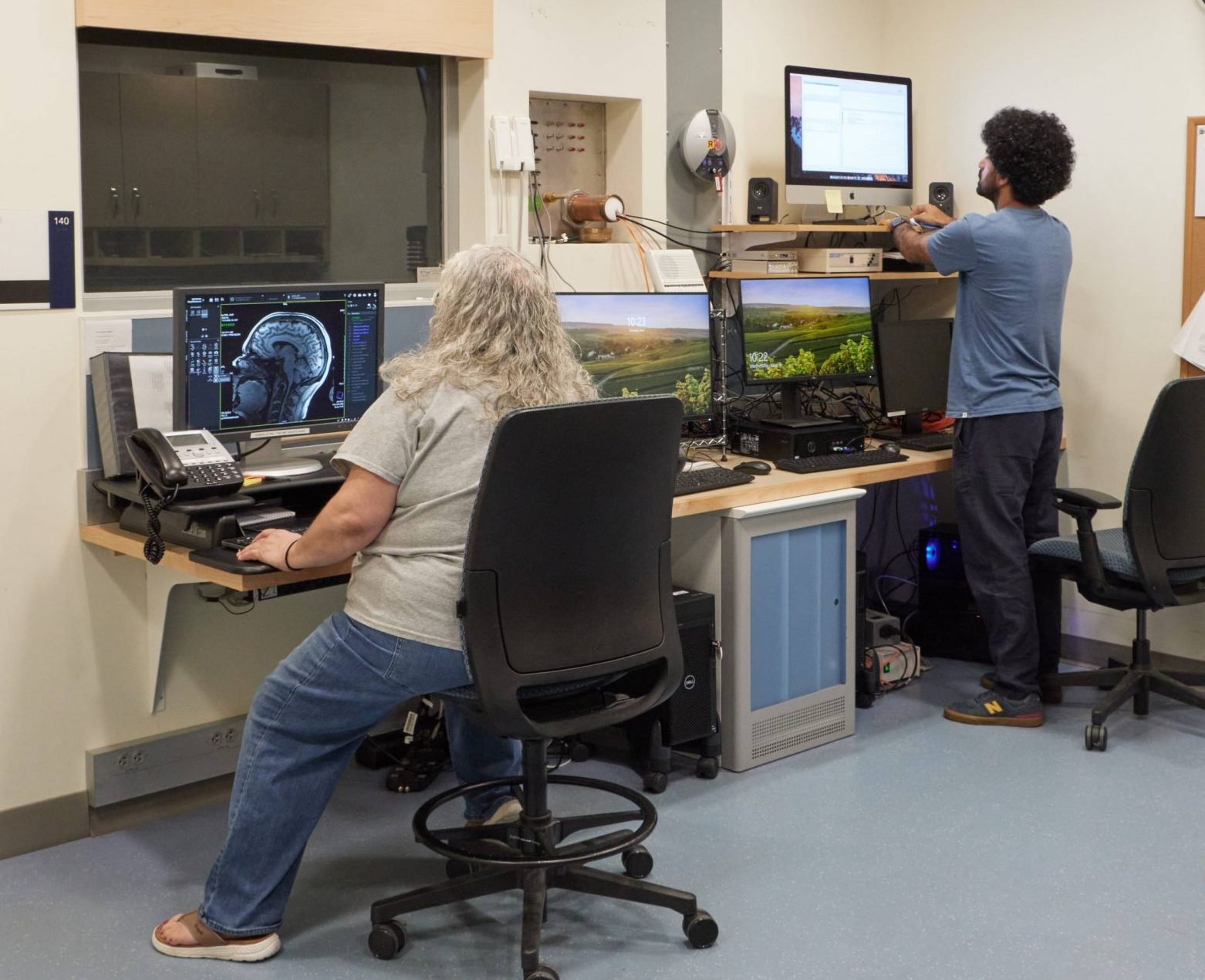
Computing
The center houses a high-end data processing lab, featuring 4 high-end Mac Workstations with common software (e.g. Docker, AFNI, FSL, Freesurfer, MATLAB, R). In addition to in-house computing, two high performance computing (HPC) systems are available to UConn faculty, providing access to accelerated GPU and parallel computing. BIRC has purchased one semi-dedicated 32-core processing node for priority access.
High-Performance Computing (HPC) facilities
The University manages HPC systems on the Storrs campus and in Farmington at UConn Health. The systems are optimized for different computational problems, with the Storrs focusing on compute-intensive workloads and the Farmington facility on data-intensive workloads. Both provide access to accelerated GPUs, parallel computing, and 1 PB of parallel file storage and 3 PB archival storage distributed across multiple datacenters. The Farmington facility has 3,000 processor cores, and the Storrs HPC cluster currently has over 11,000 CPU cores on 400 nodes. We maintain a data transfer node with Globus software for large data transfers both within and outside of the environment. Network traffic travels over Ethernet at 10Gb per second between nodes, and file data travels over InfiniBand at 56Gb or 100Gb per second, depending on the node. All UConn researchers may use the community queues, but researchers may purchase semi-dedicated nodes for high priority access. Computational applications, including containers, are installed as needed; to date, over 200 have been made available.